

Digital Pathology

Pathology workflow, digital imaging and AI solutions on a single platform.

Comprehensive Digital Pathology and Laboratory Management

RAD-X Digital Pathology System manages your digital pathology processes end to end, bringing high-resolution imaging, secure archiving and AI-assisted analysis together in a single integrated platform.

RAD-X Pathology Information Management System delivers comprehensive digital solutions for modern laboratories streamlining workflows, accelerating processes, and enhancing efficiency. The platform features fully customizable screening and diagnostic workflows, AI-driven decision support tools, and real-time monitoring to ensure precision at every stage.

Seamless barcode tracking, automated identification and intelligent monitoring eliminate manual errors and simplify daily operations. With flexible integration and an intuitive interface, RAD-X adapts effortlessly to existing laboratory infrastructures. Dynamic, live reporting provides actionable insights, while built-in quality management ensures consistent accuracy and performance.

The Entire Pathology Workflow, on One Platform

The slide is tracked by barcode in the laboratory, digitized in the scanner and examined in the viewer with AI support. RAD-X brings every step together in the same case.

Laboratory

  1. Sample Acceptance
  2. Macroscopy
  3. Tissue processing
  4. Paraffin embedding
  5. Sectioning / smear check
  6. Staining
  • Whole Slide Imaging
  • Color Calibration
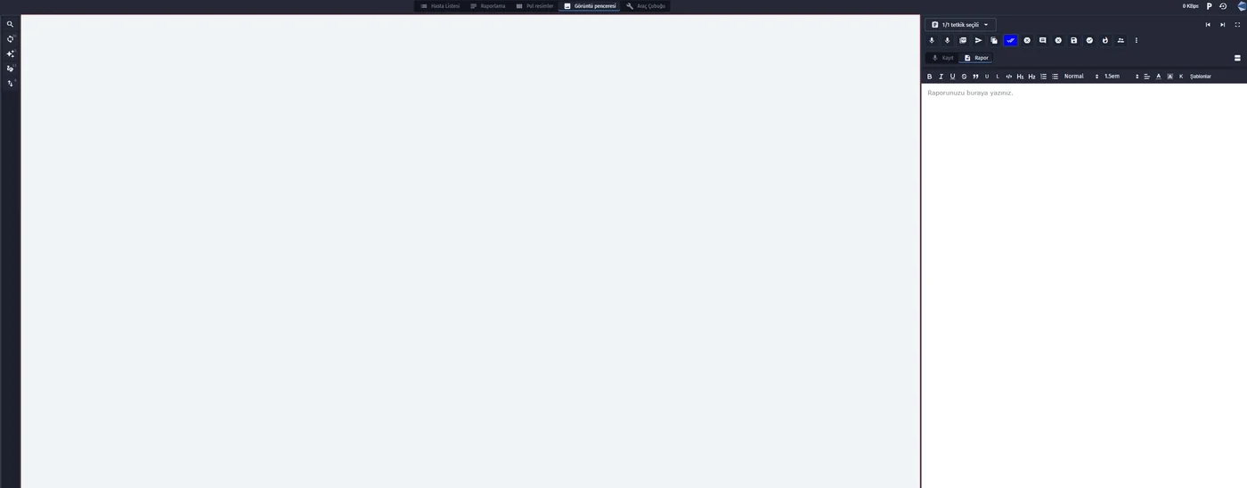
  • Multipurpose Viewer
  • Annotation Tools
  • AI Analysis
  1. Reporting
  2. Secure Archiving

Built for Hands-Free Work

RAD-X is a laboratory management system for pathology labs that keeps keyboard and mouse use to a minimum and puts ease of use and an uninterrupted workflow at its centre.

Hands-free stations

An action is chosen once per shift; from then on every scan performs it. Paraffin, sectioning, staining and archiving all run on the scanner.

Audible confirmation

Every scan answers with a sound: success, already done, or wrong item. Staff do not need to look at the screen.

No mix-ups

Cassette–slide and sample–slide matching catch a wrong slide at the bench, before it reaches the pathologist.

The case moves itself

When the last cassette or slide of a case is scanned, the case passes to the next stage automatically. Each screen shows only what is allowed at that stage.

Built from the order

Samples, cassettes and slides are created automatically according to the case type, with labels ready to print.

Full traceability

Who did what and when is recorded for every case, down to each cassette and slide, through archive, transfer and delivery.

Your Workflow, Your Rules

The workflow shown here is one configuration, not a fixed program. Stations, statuses, buttons and who may press them are data. Each laboratory shapes them to the way it works, without changing code.

Stations

Add, rename or remove a step

Each stage is a status record. A laboratory without a separate tissue processing step, or with an extra quality check, simply has a different list of stations.

Buttons

The right action at the right stage

Every action carries a rule: in which status it appears, for which case type, and which status it moves the case to. Change the rule and the screen follows.

People

By role, unit and institution

The paraffin technician sees paraffin actions, the pathologist sees reporting. Permissions are set per group and per hospital, and a rule can limit an action to the person who started the step.

Reports

Report sections you define

Macroscopy, microscopy, cytology, frozen section comment: the sections, their order and when they appear are configured per pathology type and user.

Create Your Own Scenarios

Three example cases show, step by step, how a case moves through the units of the laboratory.

From Sample Acceptance to Approved Reporting

One biopsy case with one sample, three cassettes and three slides passes through every unit of the laboratory. Apart from opening the case and approving the report, every step is done with the scanner.

  1. Macroscopy start and completion, with who and when recorded
  2. Paraffin: the action is chosen once, then each cassette is scanned
  3. Sectioning: each cassette is matched with its slide, confirmed by sound
  4. Staining completed slide by slide; the case advances by itself
  5. Sent to microscopy by barcode, blocked if any slide is unstained
  6. Reporting in defined sections and approval by the pathologist

Fast reporting for cytology cases

The system knows the case is cytology, so macroscopy, tissue processing, paraffin embedding and sectioning are skipped. The sample goes from acceptance straight to staining and microscopy, and the report is approved sooner.

  1. Sample acceptance
  2. Sample–slide verification with the scanner; a slide from another patient is rejected
  3. Smear staining started and completed by scanning
  4. Microscopy, cytology report sections and approval

Additional Stain Orders and Process Tracking

During microscopic examination or reporting, the pathologist orders a new stain. New slides are created from the order and the case goes back through sectioning and staining, then returns to the pathologist.

  1. Adding a staining order from the case screen
  2. Slides generated automatically from the order
  3. Sectioning and staining of the new slides only
  4. Back to reporting

Advanced Digital Pathology Imaging, Archiving, and AI Solutions

Multipurpose Viewer

This purpose-built viewer securely processes, stores, and manages full-section images from multiple scanners and file types (SVS, NDPI, MRXS, TIFF, DICOM, ISYNTAX), supporting multi-modality imaging and advanced hanging protocols.

Whole Slide Imaging (WSI)

Entire glass slides are scanned at high resolution, allowing pathologists to zoom in and out for detailed analysis.

Multi-level Magnification

Simulates different objective magnifications used in microscopy.

AI Integration

Supports detection, segmentation, counting, and classification of cancerous and non-cancerous cells with advanced algorithms.

Color Calibration and Standardization

Ensures consistency of images from different scanners and staining methods.

Annotation Tools

Pathologists can highlight specific regions, add notes, and share them for education or collaboration.

Remote Access (Telepathology)

Provides secure online access to experts in different locations for collaborative and remote reporting.

Archiving and Management

Enables secure long-term storage of slides with fast and easy retrieval.

AI Solutions

  • Advanced image analysis tools for detection, counting, and localization of cancerous and non-cancerous cells on ER/PR, Ki-67, HER2, and PD-L1 stained digital slides.
  • AI models to aid detection of lymph node metastasis and prostate cancer.
  • Semi-automatic cell detection based on user defined annotations.

Ki-67 Analysis · HER2 Analysis · ER/PR Analysis

Let’s Map Your Lab Workflow Together

Tell us about your stations and rules, and we’ll set RAD-X up around them.